Page 363 - Read Online
P. 363
Page 6 of 16                         Rizzieri et al. J Cancer Metastasis Treat 2019;5:26  I  http://dx.doi.org/10.20517/2394-4722.2019.05


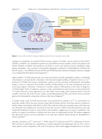
               Figure 1. Summary of the metabolic changes in multiple myeloma cells and in resistant myeloma cells

               endogenous metabolites in peripheral blood plasma samples of healthy controls, patients with MGUS,
               NDMM, or RRMM. The metabolomic profile was quite different between healthy controls and patients with
               MGUS, NDMM or RRMM with significant alterations in amino acid, lipid and energy metabolism. Eight
               plasma metabolites - free carnitine, acetylcarnitine, glutamate, asymmetric dimethylarginine (ADMA) and
               four PC species - were different between MGUS and NDMM patients, demonstrating that metabolic changes
                                                       [34]
               occur during MM development and progression .
                             1
               High-resolution  H-NMR spectroscopy and mass spectrometry provides quantitative analysis of metabolite
                                                                                                        [35]
               concentrations and reproducible information with minimal sample handling. Puchades-Carrasco et al.
                         1
               performed  H-NMR spectroscopy metabolomic analyses of serum samples of MM patients and healthy
               subjects, and also compared the metabolic profiles of MM patients at the time of diagnosis and after
               achieving complete remission. Compared to healthy subjects, MM patients at the time of diagnosis
               exhibited higher levels of isoleucine, arginine, acetate, phenylalanine, and tyrosine and decreased levels
               of 3-hydroxybutyrate, lysine, glutamine, and some lipids. Interestingly, when myeloma patients achieved
               complete remission, the levels of lysine, citrate, lactate dehydrogenase (LDH), and choline went up whereas
                                                                          [35]
               glucose level decreased in comparison to those at the time of diagnosis .
                                    1
                                                                                      [36]
               A similar study using  H-NMR spectroscopy was performed by Ludwig et al.  to characterize the
               metabolic profile within the bone marrow using filtered plasma derived from bone marrow aspirates of
               healthy donors and patients with MGUS or MM. This study found that the essential amino acids isoleucine
               and threonine were significantly decreased in the bone marrow of MGUS and MM patients as were the
               nucleotide-breakdown products, hypoxanthine and xanthine, suggesting an increase in anabolism. The
               products of arginine metabolism, creatinine and urea, were also significantly altered. The authors suggested
               that alterations in bone marrow metabolism are an early and consistent feature during the development of
                             [36]
               MGUS and MM .
                                                                 13
                                                                                   12
                                     [37]
                                                 13
               Recently, Gonsalves et al.  performed  C SIRM using U[ C ]-Glucose and U[ C ]-Glutamine in human
                                                                                      5
                                                                    6
               MM cell lines to determine the contribution of carbon substrates from glutamine into the TCA cycle of
               clonal plasma cells in comparison with those from glucose. Their results suggest that glutamine is the main

