
Page 180 - Read Online
P. 180
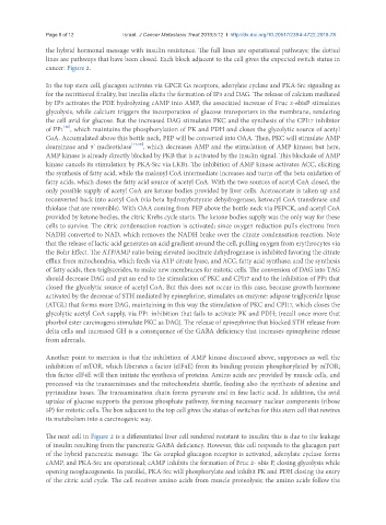
Page 8 of 12                                   Israël. J Cancer Metastasis Treat 2019;5:12  I  http://dx.doi.org/10.20517/2394-4722.2018.78

               the hybrid hormonal message with insulin resistance. The full lines are operational pathways; the dotted
               lines are pathways that have been closed. Each block adjacent to the cell gives the expected switch status in
               cancer: Figure 2.

               In the top stem cell, glucagon activates via GPCR Gs receptors, adenylate cyclase and PKA-Src signaling as
               for the nutritional finality, but insulin elicits the formation of IP3 and DAG. The release of calcium mediated
               by IP3 activates the PDE hydrolyzing cAMP into AMP, the associated increase of Fruc 2-6bisP stimulates
               glycolysis, while calcium triggers the incorporation of glucose transporters in the membrane, rendering
               the cell avid for glucose. But the increased DAG stimulates PKC and the synthesis of the CPI17 inhibitor
                     [16]
               of PP1 , which maintains the phosphorylation of PK and PDH and closes the glycolytic source of acetyl
               CoA. Accumulated above this bottle neck, PEP will be converted into OAA. Then, PKC will stimulate AMP
               deaminase and 5’ nucleotidase [17,18] , which decreases AMP and the stimulation of AMP kinase; but here,
               AMP kinase is already directly blocked by PKB that is activated by the insulin signal. This blockade of AMP
               kinase cancels its stimulation by PKA-Src via LKB1. The inhibition of AMP kinase activates ACC, eliciting
               the synthesis of fatty acid, while the malonyl CoA intermediate increases and turns off the beta oxidation of
               fatty acids, which closes the fatty acid source of acetyl CoA. With the two sources of acetyl CoA closed, the
               only possible supply of acetyl CoA are ketone bodies provided by liver cells. Acetoacetate is taken up and
               reconverted back into acetyl CoA (via beta hydroxybutyrate dehydrogenase, ketoacyl CoA transferase and
               thiolase that are reversible). With OAA coming from PEP above the bottle neck via PEPCK, and acetyl CoA
               provided by ketone bodies, the citric Krebs cycle starts. The ketone bodies supply was the only way for these
               cells to survive. The citric condensation reaction is activated; since oxygen reduction pulls electrons from
               NADH converted to NAD, which removes the NADH brake over the citrate condensation reaction. Note
               that the release of lactic acid generates an acid gradient around the cell, pulling oxygen from erythrocytes via
               the Bohr Effect. The ATP/AMP ratio being elevated isocitrate dehydrogenase is inhibited favoring the citrate
               efflux from mitochondria, which feeds via ATP citrate lyase, and ACC, fatty acid synthase; and the synthesis
               of fatty acids, then triglycerides, to make new membranes for mitotic cells. The conversion of DAG into TAG
               should decrease DAG and put an end to the stimulation of PKC and CPI17 and to the inhibition of PP1 that
               closed the glycolytic source of acetyl CoA. But this does not occur in this case, because growth hormone
               activated by the decrease of STH mediated by epinephrine, stimulates an enzyme: adipose triglyceride lipase
               (ATGL) that forms more DAG, maintaining in this way the stimulation of PKC and CPI17, which closes the
               glycolytic acetyl CoA supply, via PP1 inhibition that fails to activate PK and PDH; (recall once more that
               phorbol ester carcinogens stimulate PKC as DAG). The release of epinephrine that blocked STH release from
               delta cells and increased GH is a consequence of the GABA deficiency that increases epinephrine release
               from adrenals.


               Another point to mention is that the inhibition of AMP kinase discussed above, suppresses as well the
               inhibition of mTOR, which liberates a factor (eIF4E) from its binding protein phosphorylated by mTOR;
               this factor eIF4E will then initiate the synthesis of proteins. Amino acids are provided by muscle cells, and
               processed via the transaminases and the mitochondria shuttle, feeding also the synthesis of adenine and
               pyrimidine bases. The transamination chain forms pyruvate and in fine lactic acid. In addition, the avid
               uptake of glucose supports the pentose phosphate pathway, forming necessary nuclear components (ribose
               5P) for mitotic cells. The box adjacent to the top cell gives the status of switches for this stem cell that rewires
               its metabolism into a carcinogenic way.

               The next cell in Figure 2 is a differentiated liver cell rendered resistant to insulin; this is due to the leakage
               of insulin resulting from the pancreatic GABA deficiency. However, this cell responds to the glucagon part
               of the hybrid pancreatic message. The Gs coupled glucagon receptor is activated, adenylate cyclase forms
               cAMP, and PKA-Src are operational; cAMP inhibits the formation of Fruc 2- 6bis P, closing glycolysis while
               opening neoglucogenesis. In parallel, PKA-Src will phosphorylate and inhibit PK and PDH closing the entry
               of the citric acid cycle. The cell receives amino acids from muscle proteolysis; the amino acids follow the