
Page 81 - Read Online
P. 81
Fontes-Sousa et al. J Cancer Metastasis Treat 2018;4:5 I http://dx.doi.org/10.20517/2394-4722.2017.70 Page 3 of 6
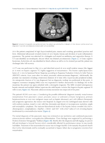
Figure 2. Computed tomography scan performed when the patient was admitted for cholangitis. A liver abscess (yellow arrow) was
diagnosed. It was later percutaneously drained and E. coli was identified
2016 the patient complained of right hypochondrial pain, nausea and vomiting, generalized pruritus and
fever. Abdominal ultrasound revealed evident de novo hepatic lesions and elevation of acute inflammatory
parameters. The patient was admitted for cholangitis and started on antibiotics and supportive therapy. A
CT scan identified an intrahepatic abscess which was drained percutaneously [Figure 2]. A Gram-negative
bacterium, Escherichia coli, was identified in blood cultures as well as in the drained pus and the patient was
discharged after full recovery.
A CT scan was performed in May 2016, and identified several de novo small nodular masses (the biggest
at 18 mm) on hepatic segment VI, highly suggestive of metastization. The known cephalopancreatic
lesion of 15 mm [a Sustained Partial Response according to Response Evaluation Criteria In Solid Tumors
(RECIST) criteria, three years after the initial pancreatic adenocarcinoma diagnosis]. Additionally, the
prostate was measured, revealing a transversal diameter of 50 mm with a hypervascular peripheral nodule.
An osteoporotic fracture of L3 was diagnosed, but no diagnostic workup was performed at this point. A
magnetic resonance imaging (MRI) scan was requested to further characterize the findings, but in the
meantime the patient was again admitted with a cholangitis diagnosis. The MRI revealed heterogeneous
hepatic steatosis and multiple bilobar hypervascular solid hepatic nodules (the largest at hepatic segment VI
with 26 mm, Figure 3A). Pancreatic adenocarcinoma metastasis was suspected at this point.
The patient’s ECOG score was 0. Considering the possible differential diagnosis (namely metachronous
metastization from pancreatic cancer, prostatic cancer or unknown primary malignancy, primary
hepatocarcinoma or even non-malignant causes such as hepatic abscesses), each with different therapeutic
and prognostic approaches, the lesions were biopsied. In August 2016 the histological exam showed cells
with uniform nucleus, round to oval, with fine chromatin and absent or inconspicuous nucleolus; ample
and eosinophilic cytoplasm. The immune-histochemical study of the hepatic lesions revealed AE1/AE3+,
chromogranin+, synaptophysin+, HepPar1-, CK903- and CK7- with a Ki67 index of 14% revealing hepatic
involvement by a neuroendocrine tumor [Figure 4].
The initial diagnosis of the pancreatic mass was reviewed at our institution and confirmed pancreatic
adenocarcinoma without neuroendocrine differentiation. These findings were supported by performing a
68
Positron Emission Tomography Gallium [Figure 3B]. Shortly after the diagnosis and full staging, the patient
fell and developed a femoral fracture which considerably affected his performance status; he was mostly
bedridden. Treatment of the neuroendocrine tumor was no longer feasible. Unfortunately, the patient’s health
then further deteriorated, and he eventually succumbed to hepatic failure due to progressive extensive liver
metastization.