
Page 23 - Read Online
P. 23
Page 4 of 24 Tokuyasu et al. J Cancer Metastasis Treat 2018;4:2 I http://dx.doi.org/10.20517/2394-4722.2017.52
All nucleated cells APC
MHC I MHC II
Peptide (8-12 Peptide (12-25
amino acid amino acid
residues) residues)
TCR TCR
CD8 + CD4 +
T cell T cell
Activation Activation
+
CD4 T
CTLs helper cells
+
+
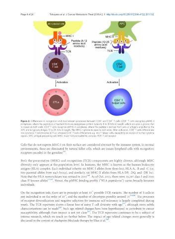
Figure 2. Differences in recognition and downstream processes between CD8 and CD4 T cells. CD8 T cells recognize pMHC-I
+
complexes, where the peptide is a fragment from an endogenous protein typically 8 to 11 AAs in length, which occupies a groove that
+
is closed on both ends. CD4 T cells recognize pMHC-II complexes, where the peptide is derived from cells or antigens engulfed by the
APC and is typically longer, 12 to 25 AAs in length. The MHC-II groove is open on both ends. After activation, CD8 T cells differentiate
+
+
into cytotoxic T lymphocytes (CTLs), whereas CD4 T cells differentiate e.g. into T helper cells, depending on receipt of further cytokine
signals. APC: antigen-presenting cell; MHC: major histocompatibility complex; TCR: T cell receptor
Cells that do not express MHC-I on their surface are considered aberrant by the immune system. In normal
environments, these are eliminated by natural killer cells, which are innate lymphoid cells with recognition
[15]
receptors encoded in the germline .
Both the presentation (MHC) and recognition (TCR) components are highly diverse, although MHC
diversity only appears at the population level. In humans, the MHC is known as the human leukocyte
antigen (HLA) complex. Each individual inherits six MHC-I alleles from three loci, HLA-A, -B and -C (i.e.
two parental alleles from each locus), and similarly, six MHC-II alleles from HLA-DP, -DQ, and -DR loci.
[16]
Note that the HLA nomenclature was revised in 2010 . As of Oct. 2015, there were 10,297 class I and 3543
class II known alleles [17,18] . Hence, the pMHC binding profile (“HLA peptidome”) varies broadly between
individuals.
15
On the recognition side, there are in principle at least 10 possible TCR variants. The number of T-cells in
12
any individual is on the order of 10 , and the number of clonotypes possibly around 10 7[19,20] . The processes
of receptor diversification and negative selection for immune self-tolerance is largely completed during
[21]
youth. The TCR repertoire shows a linear loss of naive T cell diversity with age , although more subtle
[22]
characterizations can be made . Such age-related changes have been hypothesized to contribute to cancer
[23]
susceptibility, although their impact is not yet clear . The TCR repertoire continues to be a subject of
intense research, which we touch on further below. The impact of age-related changes more generally is
[24]
discussed in the context of checkpoint blockade therapy by Elias et al. .