
Page 67 - Read Online
P. 67
Page 4 of 22 García-Pardo et al. J Cancer Metastasis Treat 2021;7:62 https://dx.doi.org/10.20517/2394-4722.2021.103
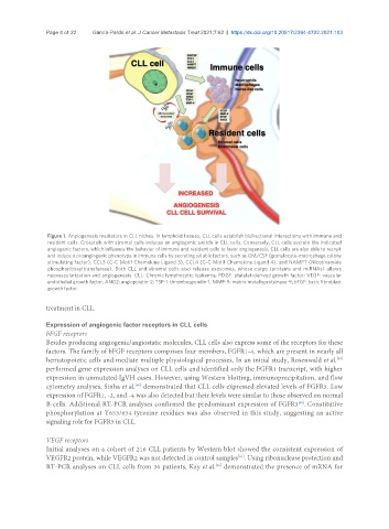
Figure 1. Angiogenesis mediators in CLL niches. In lymphoid tissues, CLL cells establish bidirectional interactions with immune and
resident cells. Crosstalk with stromal cells induces an angiogenic switch in CLL cells. Conversely, CLL cells secrete the indicated
angiogenic factors, which influence the behavior of immune and resident cells to favor angiogenesis. CLL cells are also able to recruit
and induce a proangiogenic phenotype in immune cells by secreting soluble factors, such as GM/CSF (granulocyte-macrophage colony
stimulating factor), CCL3 (C-C Motif Chemokine Ligand 3), CCL4 (C-C Motif Chemokine Ligand 4), and NAMPT (Nicotinamide
phosphoribosyltransferase). Both CLL and stromal cells also release exosomes, whose cargo (proteins and miRNAs) allows
neovascularization and angiogenesis. CLL: Chronic lymphocytic leukemia; PDGF: platelet-derived growth factor; VEGF: vascular
endothelial growth factor; ANG2: angiopoietin-2; TSP-1: thrombospondin-1; MMP-9: matrix metalloproteinase-9; bFGF: basic fibroblast
growth factor.
treatment in CLL.
Expression of angiogenic factor receptors in CLL cells
bFGF receptors
Besides producing angiogenic/angiostatic molecules, CLL cells also express some of the receptors for these
factors. The family of bFGF receptors comprises four members, FGFR1-4, which are present in nearly all
hematopoietic cells and mediate multiple physiological processes. In an initial study, Rosenwald et al.
[59]
performed gene expression analyses on CLL cells and identified only the FGFR1 transcript, with higher
expression in unmutated IgVH cases. However, using Western blotting, immunoprecipitation, and flow
cytometry analyses, Sinha et al. demonstrated that CLL cells expressed elevated levels of FGFR3. Low
[60]
expression of FGFR1, -2, and -4 was also detected but their levels were similar to those observed on normal
B-cells. Additional RT-PCR analyses confirmed the predominant expression of FGFR3 . Constitutive
[60]
phosphorylation at Y653/654 tyrosine residues was also observed in this study, suggesting an active
signaling role for FGFR3 in CLL.
VEGF receptors
Initial analyses on a cohort of 216 CLL patients by Western blot showed the consistent expression of
VEGFR2 protein, while VEGFR2 was not detected in control samples . Using ribonuclease protection and
[61]
RT-PCR analyses on CLL cells from 30 patients, Kay et al. demonstrated the presence of mRNA for
[36]