
Page 390 - Read Online
P. 390
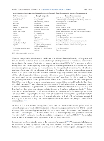
Darbre. J Cancer Metastasis Treat 2019;5:58  I  http://dx.doi.org/10.20517/2394-4722.2019.22                                   Page 3 of 13

               Table 1. Estrogen disrupting chemicals: example compounds, uses in the environment, and sources of human exposure
               Use in the environment     Example compounds                Source of human exposure
               Plant phytoestrogens     Genistein, daidzein      Edible plant material (diet, dietary supplements)
               Pharmaceuticals          Ethinylestradiol         Contraceptive pill, hormone replacement therapy, cosmetics
               Pesticides               DDT, lindane, pyrethroids  Pesticide products, animal fat (diet)
               Herbicides               Glyphosate               Garden weedkillers
               Industrial               Polychlorinated biphenyls  Animal fat (diet)
               By-product of incineration  Polychlorinated dioxins  Inhaled; animal fat in diet
               Flame retardant          Polybrominated organics  Domestic environment from use on soft furnishings
               Plastics, epoxy resins   Bisphenol A              Storage of food and beverages (diet); domestic environment
               Plastics                 Phthalate esters         Domestic consumer products
               Detergent                Alkyl phenols            Domestic environment
               Preservative             Parabens                 Personal care products, food, pharmaceuticals
               Antiperspirant           Aluminium chlorohydrate  Underarm antiperspirants
               Antimicrobial            Triclosan                Personal care products, domestic consumer products
               Absorb ultraviolet light  Benzophenones           Suncare products, other cosmetics, clothing
               Fragrance                Butylphenylmethylpropional,   Personal care products, domestic consumer products
                                        benzyl salicylate, musks
               Conditioning/spreading agent  Cyclic volatile methylsiloxanes  Personal care products
               Cigarettes               Cadmium                  Cigarette smoking

               DDT: dichlorodiphenyltrichloroethane

               However, endogenous estrogens are also now known to be able to influence cell motility, cell migration and
               invasive behavior of human breast cancer cells through altering expression of proteins and transcription
               factors key to the process of epithelial to mesenchymal transition (EMT). EMT is a process in which
               the epithelial cells lose their polarity and strong cell-cell adhesion properties in order to assume a more
               mesenchymal phenotype lacking the polarization and the strong cell-cell interactions. The cell adhesion
               junctions of epithelial cells are reliant on high levels of E-cadherin which is a transmembrane protein
               linked to the cytoskeleton by a- and b-catenin, and EMT is typically associated with reduction in levels
               of these adhesion proteins. It is also associated with altered levels of transcription factors (such as slug
                                                                    [7]
               and snail) which control expression of the adhesion proteins . This allows the cells to break away from
               neighbouring cells and to become generally more motile. Human breast cancer cell lines which possess
               ERa (ERa+) tend to be less invasive, less metastatic and possess higher levels of E-cadherin than those
                                   [11]
               which lack ERa (ERa-) . Knockdown of ERa in the ERa+ cell lines has been shown to result in decreased
                                                  [12]
               E-cadherin and increased slug expression . Conversely, transfected overexpression of ERa in the ERa- cell
               lines has been shown to enable estrogen-mediated increase in E-cadherin and decrease in slug . In the
                                                                                                  [12]
               ERa+ MCF-7 human breast cancer cell line, estradiol can increase EMT whilst the antiestrogen tamoxifen
               can reduce EMT  suggesting that the mechanism is ER-dependent. However, there appear to be multiple
                             [13]
               molecular mechanisms involving not only genomic alterations to levels of E-cadherin and associated
                                           [12]
               transcription factors such as slug  but also non-genomic alterations via c-src and phosphorylation of focal
               adhesion kinase [14,15] .

               In order to facilitate invasion through local tissue, the cells need also to secrete greater levels of
               extracellular proteases which aid in the digestion of the surrounding extracellular matrix (ECM). Some of
               the extracellular proteases identified specifically in human breast cancer cells are matrix metalloproteinases
                                        [18]
               (MMPs) [16,17]  and cathepsin D . It is notable that one of the first estrogen-regulated genes to be identified
                                                                                            [16]
                             [19]
               was cathepsin D  and studies now also show effects of estrogen on expression of MMPs . These studies
               attest to the role of estrogen in altering proteases which can degrade the ECM.
               Estrogens are also known to play a role in the processes of angiogenesis [10,20] . Vascular endothelial growth
               factor (VEGF) is a key angiogenesis promoting factor and the VEGF gene has been shown to possess
               functional estrogen response elements indicating it is an estrogen-responsive gene . Estrogen treatment
                                                                                      [21]