
Page 126 - Read Online
P. 126
Saccà et al. J Cancer Metastasis Treat 2019;5:15 I http://dx.doi.org/10.20517/2394-4722.2018.95 Page 5 of 9
A B
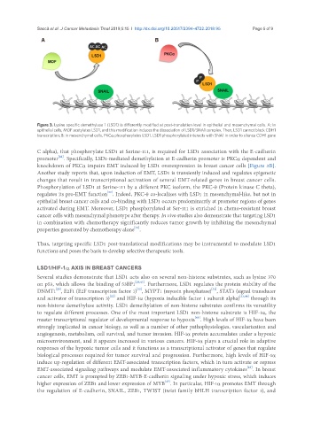
Figure 3. Lysine specific demethylase 1 (LSD1) is differently modified at post-translation level in epithelial and mesenchymal cells. A: In
epithelial cells, MOF acetylates LSD1, and this modification induces the dissociation of LSD1/SNA1 complex. Then, LSD1 cannot block CDH1
transcription; B: in mesenchymal cells, PKCα phosphorylates LSD1. LSD1 phosphorylated interacts with SNA1 in order to silence CDH1 gene
C alpha), that phosphorylate LSD1 at Serine-111, is required for LSD1 association with the E-cadherin
[42]
promoter . Specifically, LSD1-mediated demethylation at E-cadherin promoter is PKCα dependent and
knockdown of PKCα impairs EMT induced by LSD1 overexpression in breast cancer cells [Figure 3B].
Another study reports that, upon induction of EMT, LSD1 is transiently induced and regulates epigenetic
changes that result in transcriptional activation of several EMT-related genes in breast cancer cells.
Phosphorylation of LSD1 at Serine-111 by a different PKC isoform, the PKC-θ (Protein kinase C theta),
[22]
regulates its pro-EMT function . Indeed, PKC-θ co-localizes with LSD1 in mesenchymal-like, but not in
epithelial breast cancer cells and co-binding with LSD1 occurs predominantly at promoter regions of genes
activated during EMT. Moreover, LSD1 phosphorylated at Ser-111 is enriched in chemo-resistant breast
cancer cells with mesenchymal phenotype after therapy. In vivo studies also demonstrate that targeting LSD1
in combination with chemotherapy significantly reduces tumor growth by inhibiting the mesenchymal
[22]
properties generated by chemotherapy alone .
Thus, targeting specific LSD1 post-translational modifications may be instrumental to modulate LSD1
functions and poses the basis to develop selective therapeutic tools.
LSD1/HIF-1α AXIS IN BREAST CANCERS
Several studies demonstrate that LSD1 acts also on several non-histone substrates, such as lysine 370
on p53, which allows the binding of 53BP1 [28,43] . Furthermore, LSD1 regulates the protein stability of the
[29]
[31]
[30]
DNMT1 , E2F1 (E2F transcription factor 1) , MYPT1 (myosin phosphatase) , STAT3 (signal transducer
[32]
and activator of transcription 3) and HIF-1α (hypoxia inducible factor 1 subunit alpha) [33,44] through its
non-histone demethylase activity. LSD1 demethylation of non-histone substrates confirms its versatility
to regulate different processes. One of the most important LSD1 non-histone substrate is HIF-1α, the
[45]
master transcriptional regulator of developmental response to hypoxia . High levels of HIF-1α have been
strongly implicated in cancer biology, as well as a number of other pathophysiologies, vascularization and
angiogenesis, metabolism, cell survival, and tumor invasion. HIF-1α protein accumulates under a hypoxic
microenvironment, and it appears increased in various cancers. HIF-1α plays a crucial role in adaptive
responses of the hypoxic tumor cells and it functions as a transcriptional activator of genes that regulate
biological processes required for tumor survival and progression. Furthermore, high levels of HIF-1α
induce up-regulation of different EMT-associated transcription factors, which in turn activate or repress
[46]
EMT-associated signaling pathways and modulate EMT-associated inflammatory cytokines . In breast
cancer cells, EMT is prompted by ZEB1-MYB-E-cadherin signaling under hypoxic stress, which induces
[47]
higher expression of ZEB1 and lower expression of MYB . In particular, HIF-1α promotes EMT through
the regulation of E-cadherin, SNAIL, ZEB1, TWIST (twist family bHLH transcription factor 1), and