
Page 65 - Read Online
P. 65
Tokuyasu et al. J Cancer Metastasis Treat 2018;4:2 I http://dx.doi.org/10.20517/2394-4722.2017.52 Page 3 of 24
All nucleated cells
APC
MHC II
MHC I
CD80/86 CD80/86
CD28 TCR CD28 TCR
CD4 +
CD8 + T cell
Proliferation, Proliferation,
Differentiation, Differentiation,
Cytokine secretion Cytokine secretion
+
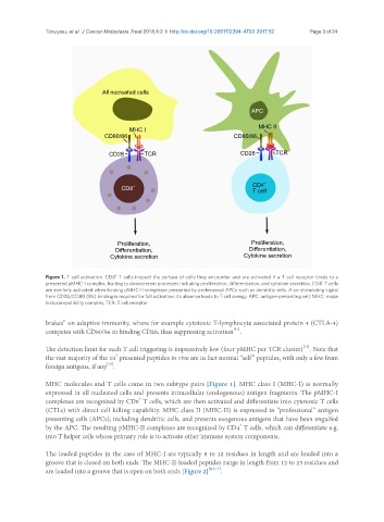
Figure 1. T cell activation. CD8 T cells inspect the surface of cells they encounter and are activated if a T cell receptor binds to a
+
presented pMHC-I complex, leading to downstream processes including proliferation, differentiation, and cytokine secretion. CD4 T cells
are similarly activated when binding pMHC-II complexes presented by professional APCs such as dendritic cells. A co-stimulating signal
from CD28/CD80 (86) binding is required for full activation; its absence leads to T cell anergy. APC: antigen-presenting cell; MHC: major
histocompatibility complex; TCR: T cell receptor
brakes” on adaptive immunity, where for example cytotoxic T-lymphocyte associated protein 4 (CTLA-4)
[11]
competes with CD80/86 in binding CD28, thus suppressing activation .
[12]
The detection limit for such T cell triggering is impressively low (four pMHC per TCR cluster) . Note that
4
the vast majority of the 10 presented peptides in vivo are in fact normal “self” peptides, with only a few from
[13]
foreign antigens, if any .
MHC molecules and T cells come in two subtype pairs [Figure 1]. MHC class I (MHC-I) is normally
expressed in all nucleated cells and presents intracellular (endogenous) antigen fragments. The pMHC-I
+
complexes are recognized by CD8 T cells, which are then activated and differentiate into cytotoxic T cells
(CTLs) with direct cell killing capability. MHC class II (MHC-II) is expressed in “professional” antigen
presenting cells (APCs), including dendritic cells, and presents exogenous antigens that have been engulfed
+
by the APC. The resulting pMHC-II complexes are recognized by CD4 T cells, which can differentiate e.g.
into T helper cells whose primary role is to activate other immune system components.
The loaded peptides in the case of MHC-I are typically 8 to 12 residues in length and are loaded into a
groove that is closed on both ends. The MHC-II-loaded peptides range in length from 12 to 25 residues and
are loaded into a groove that is open on both ends [Figure 2] [6,7,14] .