
Page 983 - Read Online
P. 983
Page 4 of 18 Machado. Hepatoma Res 2020;6:84 I http://dx.doi.org/10.20517/2394-5079.2020.90
Figure 1. Pathogenesis of lean-NAFLD. NAFLD: nonalcoholic fatty liver disease
More important than BMI, a poor surrogate for adiposity, is central obesity which seems to predispose
individuals to lean-NAFLD. In fact, lean patients with NAFLD tend to have higher WC as compared to
[32]
lean subjects without NAFLD . When comparing lean NAFLD with obese NAFLD, expectedly lean
subjects present with lower WC. However, lean patients with NAFLD present similar waist-to-hip (WTH)
[20]
[22]
ratio but higher visceral adiposity indexes as compared to obese patients . This can be explained by
the poor accuracy of WC for visceral adipose tissue (VAT). In fact, WC is determined by both visceral and
[37]
abdominal subcutaneous fat .
Lastly, in lean individuals, higher ferritin and hemoglobin levels seem to be associated with NAFLD, which
suggest iron overload and oxidative stress as a cofactor to lipotoxicity in this subset of patients [32,38] .
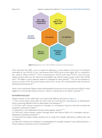
PATHOPHYSIOLOGY
Hepatic steatosis, in lean individuals, can be induced by different mechanisms [Figure 1]:
(1) The visceral adipose tissue spills over fatty acids that reach the liver, and promotes an inflammatory
milieu, producing adipokines that are diabetogenic and steatogenic ;
[39]
(2) The liver itself is prone to steatosis, for example, as a consequence of genetic traits that modulate the
[40]
metabolism and export of lipids ;
(3) Malnutrition and malabsorptive diseases induce hepatic steatosis [41,42] ;
(4) The bowel induces hepatic steatosis through direct and indirect effects of a steatogenic and pro-
[43]
inflammatory intestinal microbiota ;
(5) External factors induce hepatic steatosis such as drugs (for example, amiodarone, methotrexate, and
tamoxifen);
(6) Other liver diseases may predispose to steatohepatitis (for example, hepatitis C particularly genotype 3,
[44]
Wilson’s disease, and inborn errors of metabolism) .