
Page 585 - Read Online
P. 585
Page 4 of 11 Kanmaniraja et al. Hepatoma Res 2020;6:51 I http://dx.doi.org/10.20517/2394-5079.2020.46
Figure 1. Liver Imaging Reporting and Data System computed tomography/magnetic resonance imaging diagnostic table, used to assign
LR-3, LR-4, and LR-5 categories. Reprinted with permission from Ref. [8]
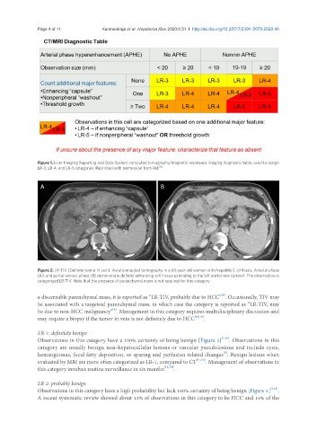
A B
Figure 2. LR-TIV (Definite tumor in vein). Axial computed tomography in a 63-year-old woman with hepatitis C cirrhosis. Arterial phase
(A); and portal venous phase (B) demonstrate definite enhancing soft tissue extending to the left portal vein (arrow). The observation is
categorized LR-TIV. Note that the presence of parenchymal mass is not required for this category
[9]
a discernable parenchymal mass, it is reported as “LR-TIV, probably due to HCC” . Occasionally, TIV may
be associated with a targetoid parenchymal mass, in which case the category is reported as “LR-TIV, may
[9]
be due to non-HCC malignancy” . Management in this category requires multidisciplinary discussion and
may require a biopsy if the tumor in vein is not definitely due to HCC [16,18] .
LR-1: definitely benign
Observations in this category have a 100% certainty of being benign [Figure 3] [7,15] . Observations in this
category are usually benign non-hepatocellular lesions or vascular pseudolesions and include cysts,
[9]
hemangiomas, focal fatty deposition, or sparing and perfusion related changes . Benign lesions when
evaluated by MRI are more often categorized as LR-1, compared to CT [11,12] . Management of observations in
this category involves routine surveillance in six months [16,18] .
LR-2: probably benign
Observations in this category have a high probability but lack 100% certainty of being benign [Figure 4] [7,15] .
A recent systematic review showed about 13% of observations in this category to be HCC and 14% of the