
Page 140 - Read Online
P. 140
Chen et al. Hepatoma Res 2019;5:12 I http://dx.doi.org/10.20517/2394-5079.2019.03 Page 3 of 17
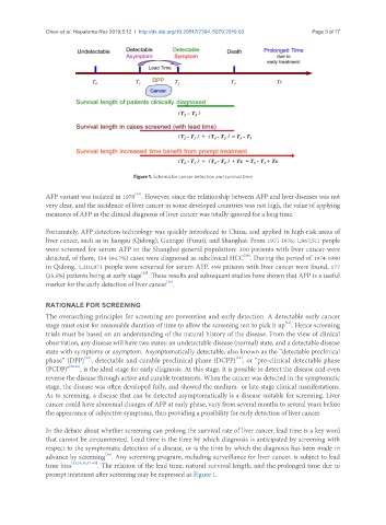
Figure 1. Schema for cancer detection and survival time
[29]
AFP variant was isolated in 1970 . However, since the relationship between AFP and liver diseases was not
very clear, and the incidence of liver cancer in some developed countries was not high, the value of applying
measures of AFP in the clinical diagnosis of liver cancer was totally ignored for a long time.
Fortunately, AFP detection technology was quickly introduced to China, and applied in high-risk areas of
liver cancer, such as in Jiangsu (Qidong), Guangxi (Fusui), and Shanghai. From 1971-1976, 1,967,511 people
were screened for serum AFP in the Shanghai general population: 300 patients with liver cancer were
detected, of them, 134 (44.7%) cases were diagnosed as subclinical HCC . During the period of 1974-1980
[30]
in Qidong, 1,310,871 people were screened for serum AFP, 499 patients with liver cancer were found, 177
[15]
(35.5%) patients being at early stage . These results and subsequent studies have shown that AFP is a useful
[16]
marker for the early detection of liver cancer .
RATIONAle fOR sCReeNINg
The overarching principles for screening are prevention and early detection. A detectable early cancer
[31]
stage must exist for reasonable duration of time to allow the screening test to pick it up . Hence screening
trials must be based on an understanding of the natural history of the disease. From the view of clinical
observation, any disease will have two states: an undetectable disease (normal) state, and a detectable disease
state with symptoms or asymptom. Asymptomatically detectable, also known as the “detectable preclinical
[32]
[33]
phase” (DPP) , detectable and curable preclinical phase (DCPP) , or “pre-clinical detectable phase
(PCDP)” [34,35] , is the ideal stage for early diagnosis. At this stage, it is possible to detect the disease and even
reverse the disease through active and curable treatments. When the cancer was detected in the symptomatic
stage, the disease was often developed fully, and showed the medium- or late-stage clinical manifestations.
As to screening, a disease that can be detected asymptomatically is a disease suitable for screening. Liver
cancer could have abnormal changes of AFP at early phase, vary from several months to several years before
the appearance of subjective symptoms, thus providing a possibility for early detection of liver cancer.
In the debate about whether screening can prolong the survival rate of liver cancer, lead time is a key word
that cannot be circumvented. Lead time is the time by which diagnosis is anticipated by screening with
respect to the symptomatic detection of a disease, or is the time by which the diagnosis has been made in
advance by screening . Any screening program, including surveillance for liver cancer, is subject to lead
[36]
time bias [23,31,35,37-40] . The relation of the lead time, natural survival length, and the prolonged time due to
prompt treatment after screening may be expressed as Figure 1.