Page 187 - Read Online
P. 187
Naeem et al. Hepatoma Res 2018;4:18  I  http://dx.doi.org/10.20517/2394-5079.2018.22                                         Page 5 of 10

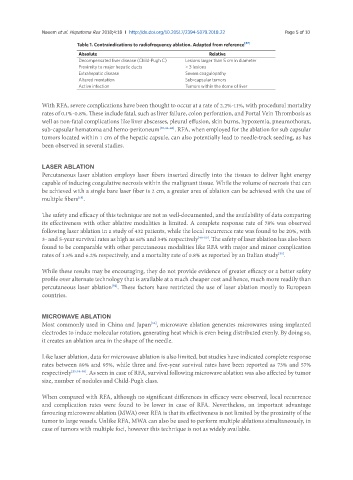
                           Table 1. Contraindications to radiofrequency ablation. Adapted from reference [47]
                            Absolute                                                    Relative
                            Decompensated liver disease (Child-Pugh C)  Lesions larger than 5 cm in diameter
                            Proximity to major hepatic ducts    > 3 lesions
                            Extrahepatic disease                Severe coagulopathy
                            Altered mentation                   Sub-capsular tumors
                            Active infection                    Tumors within the dome of liver

               With RFA, severe complications have been thought to occur at a rate of 2.2%-11%, with procedural mortality
               rates of 0.1%-0.8%. These include fatal, such as liver failure, colon perforation, and Portal Vein Thrombosis as
               well as non-fatal complications like liver abscesses, pleural effusion, skin burns, hypoxemia, pneumothorax,
               sub-capsular hematoma and hemo-peritoneum [30,44-46] . RFA, when employed for the ablation for sub capsular
               tumors located within 1 cm of the hepatic capsule, can also potentially lead to needle-track seeding, as has
               been observed in several studies.


               LASER ABLATION
               Percutaneous laser ablation employs laser fibers inserted directly into the tissues to deliver light energy
               capable of inducing coagulative necrosis within the malignant tissue. While the volume of necrosis that can
               be achieved with a single bare laser fiber is 2 cm, a greater area of ablation can be achieved with the use of
               multiple fibers .
                           [18]
               The safety and efficacy of this technique are not as well-documented, and the availability of data comparing
               its effectiveness with other ablative modalities is limited. A complete response rate of 78% was observed
               following laser ablation in a study of 432 patients, while the local recurrence rate was found to be 20%, with
               3- and 5-year survival rates as high as 61% and 34% respectively [48-50] . The safety of laser ablation has also been
               found to be comparable with other percutaneous modalities like RFA with major and minor complication
               rates of 1.5% and 6.2% respectively, and a mortality rate of 0.8% as reported by an Italian study .
                                                                                               [51]
               While these results may be encouraging, they do not provide evidence of greater efficacy or a better safety
               profile over alternate technology that is available at a much cheaper cost and hence, much more readily than
               percutaneous laser ablation . These factors have restricted the use of laser ablation mostly to European
                                       [52]
               countries.


               MICROWAVE ABLATION
               Most commonly used in China and Japan , microwave ablation generates microwaves using implanted
                                                    [53]
               electrodes to induce molecular rotation, generating heat which is even being distributed evenly. By doing so,
               it creates an ablation area in the shape of the needle.

               Like laser ablation, data for microwave ablation is also limited, but studies have indicated complete response
               rates between 89% and 95%, while three and five-year survival rates have been reported as 73% and 57%
               respectively [25,54-58] . As seen in case of RFA, survival following microwave ablation was also affected by tumor
               size, number of nodules and Child-Pugh class.

               When compared with RFA, although no significant differences in efficacy were observed, local recurrence
               and complication rates were found to be lower in case of RFA. Nevertheless, an important advantage
               favouring microwave ablation (MWA) over RFA is that its effectiveness is not limited by the proximity of the
               tumor to large vessels. Unlike RFA, MWA can also be used to perform multiple ablations simultaneously, in
               case of tumors with multiple foci, however this technique is not as widely available.

