
Page 95 - Read Online
P. 95
Page 4 of 18 Alqahtani et al. Hepatoma Res 2020;6:58 I http://dx.doi.org/10.20517/2394-5079.2020.49
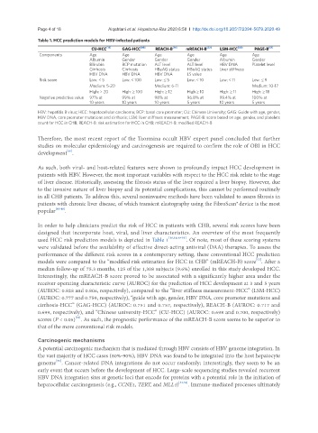
Table 1. HCC prediction models for HBV-infected patients
CU-HCC [18] GAG-HCC [49] REACH-B [16] mREACH-B [52] LSM-HCC [50] PAGE-B [51]
Components Age Age Age Age Age Age
Albumin Gender Gender Gender Albumin Gender
Bilirubin BCP mutation ALT level ALT level HBV DNA Platelet level
Cirrhosis Cirrhosis HBeAG status HBeAG status Liver stiffness
HBV DNA HBV DNA HBV DNA LS value
Risk score Low: < 5 Low: < 100 Low: ≤ 5 Low: < 10 Low: < 11 Low: ≤ 9
Medium: 5-20 Medium: 6-11 Medium: 10-17
High: > 20 High: ≥ 100 High: ≥ 12 High ≥ 10 High: ≥ 11 High: ≥ 18
Negative predictive value 97% at 99% at 98% at 96.8% at 99.4% at 100% at
10 years 10 years 10 years 5 years 10 years 5 years
HBV: hepatitis B virus; HCC: hepatocellular carcinoma; BCP: basal core promoter; CU: Chinese University; GAG: Guide with age, gender,
HBV DNA, core promoter mutations and cirrhosis; LSM: liver stiffness measurement; PAGE-B: score based on age, gender, and platelets
count for HCC in CHB; REACH-B: risk estimation for HCC in CHB; mREACH-B: modified REACH-B
Therefore, the most recent report of the Taormina occult HBV expert panel concluded that further
studies on molecular epidemiology and carcinogenesis are required to confirm the role of OBI in HCC
[40]
development .
As such, both viral- and host-related features were shown to profoundly impact HCC development in
patients with HBV. However, the most important variables with respect to the HCC risk relate to the stage
of liver disease. Historically, assessing the fibrosis status of the liver required a liver biopsy. However, due
to the invasive nature of liver biopsy and its potential complications, this cannot be performed routinely
in all CHB patients. To address this, several noninvasive methods have been validated to assess fibrosis in
patients with chronic liver disease, of which transient elastography using the FibroScan®device is the most
popular [46-48] .
In order to help clinicians predict the risk of HCC in patients with CHB, several risk scores have been
designed that incorporate host, viral, and liver characteristics. An overview of the most frequently
used HCC risk prediction models is depicted in Table 1 [16,18,49-51] . Of note, most of these scoring systems
were validated before the availability of effective direct-acting antiviral (DAA) therapies. To assess the
performance of the different risk scores in a contemporary setting, these conventional HCC prediction
models were compared to the “modified risk estimation for HCC in CHB” (mREACH-B) score . After a
[52]
median follow-up of 75.3 months, 125 of the 1,308 subjects (9.6%) enrolled in this study developed HCC.
Interestingly, the mREACH-B score proved to be associated with a significantly higher area under the
receiver operating characteristic curve (AUROC) for the prediction of HCC development at 3 and 5 years
(AUROC: 0.828 and 0.806, respectively), compared to the “liver stiffness measurement-HCC” (LSM-HCC)
(AUROC: 0.777 and 0.759, respectively), “guide with age, gender, HBV DNA, core promoter mutations and
cirrhosis-HCC” (GAG-HCC) (AUROC: 0.751 and 0.757, respectively), REACH-B (AUROC: 0.717 and
0.699, respectively), and “Chinese university-HCC” (CU-HCC) (AUROC: 0.698 and 0.700, respectively)
scores (P < 0.05) . As such, the prognostic performance of the mREACH-B score seems to be superior to
[52]
that of the more conventional risk models.
Carcinogenic mechanisms
A potential carcinogenic mechanism that is mediated through HBV consists of HBV genome integration. In
the vast majority of HCC cases (80%-90%), HBV DNA was found to be integrated into the host hepatocyte
[53]
genome . Cancer-related DNA integrations do not occur randomly; interestingly, they seem to be an
early event that occurs before the development of HCC. Large-scale sequencing studies revealed recurrent
HBV DNA integration sites at genetic loci that encode for proteins with a potential role in the initiation of
hepatocellular carcinogenesis (e.g., CCNE1, TERT, and MLL4) [53,54] . Immune-mediated processes ultimately