
Page 92 - Read Online
P. 92
Bellentani. Hepatoma Res 2020;6:29 I http://dx.doi.org/10.20517/2394-5079.2020.10 Page 3 of 6
[5]
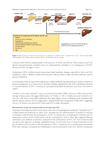
Figure 1. Natural history of NASH and risk factors for progression of NASH to HCC. Adapted from study . NASH: nonalcoholic
steatohepatitis; HCC: hepatocellular carcinoma; NAFLD: nonalcoholic fatty liver disease
of obesity with NAFLD, independently of the presence of NASH and fibrosis. Their studies reveal how
obesity-associated hepatic oxidative stress can independently contribute to the pathogenesis of NASH,
fibrosis and HCC, but again in mice.
Furthermore, HCC exhibits several characteristic lipid metabolic changes, especially in fatty acid (FA)
metabolism, which is linked to NASH and associated with the status of other metabolic pathways, such as
glucose metabolism.
Accumulating evidence supports the importance of lipid metabolic reprogramming in various situations of
hepatocarcinogenesis. In a recent review, the latest findings regarding the role of FA metabolism pathways
in the development of HCC, focusing on reprogramming lipid metabolism, have been discussed in
[17]
depth .
[18]
Another recent study reported that unconventional prefoldin RPB5 interactor (URI) induces DNA
damage in hepatocytes and triggers inflammation via T helper 17 (Th17) lymphocytes and interleukin-17A
(IL-17A). This induces neutrophil infiltration into white adipose tissue, mediating insulin resistance (IR)
and FA release, stored in liver as triglycerides, causing NASH and subsequently NASH-HCC, suggesting
that IL-17A blockers may prevent IR, NASH, and HCC in high-risk patients.
Mechanisms of gut microbiota-induced obesity and HCC
[19]
Alterations of intestinal microbiota seem to play a key role in this pathogenetic mechanism. Yoshimoto et al.
studied hepatocarcinogenesis in obese mice, showing that the administration of antibiotics and gut
sterilization could decrease the development of HCC in treated mice, modulating the dysbiosis and the
subsequent secretion of pro-inflammatory and pro-carcinogenetic factors. Their data suggested that gut
sterilization and antibiotic treatments could prevent the development of HCC, but these treatments did
not lead to the regression of already established tumors. Several other lines of research showed that the
gut microbiota is also involved in the development of HCC, in particular by increasing lipopolysaccharide
levels and creating a subsequent pro-inflammatory microenvironment in the liver. Another mechanism