
Page 24 - Read Online
P. 24
Raza et al. Hepatoma Res 2019;5:42 I http://dx.doi.org/10.20517/2394-5079.2019.014 Page 3 of 11
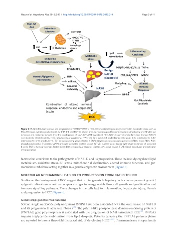
Figure 1. Multiple hits lead to onset and progression of NAFLD/NASH to HCC. Diverse signalling pathways involved in metabolic stress such as
FFAs ER-stress, cytokine production (IL-6, IL-17, IL-11 and TGF-β), altered immune response, pro-fibrogenic mediators (hedgehog and NF-κB), gut
dysbiosis and endocrine defects drive the development of NAFLD/NASH-associated HCC. NAFLD: non-alcoholic fatty liver disease; NASH:
non-alcoholic steatohepatitis; HCC: hepatocellular carcinoma; FFAs: free fatty acids; ER: endoplasmic reticulum; IL-6: interleukin-6; IL-17:
interleukin-17; IL-11: interleukin-11; TGF-β: transforming growth factor β; SNPs: single nucleotide polymorphisms; miRNA: micro RNA; PI3K:
phosphatidylinositol 3-kinases; MAPK: mitogen-activated protein kinase; NF-κB: nuclear factor kappa-light-chain-enhancer of activated
B-cells; TNF-α: tumour necrosis factor-alpha; ERK: extracellular receptor kinase; JAK: Janus kinase; STAT: signal transducer and activator
of transcription
factors that contribute to the pathogenesis of NAFLD and its progression. These include: dysregulated lipid
metabolism, oxidative stress, ER stress, mitochondrial dysfunction, altered immune function, and gut-
microbiota imbalance acting together in a genetic/epigenetic environment [Figure 1].
MOLECULAR MECHANISMS LEADING TO PROGRESSION FROM NAFLD TO HCC
Studies on the development of HCC suggest that carcinogenesis in hepatocytes is a consequence of genetic/
epigenetic alterations as well as complex changes in energy metabolism, cell growth and proliferation and
immune signalling pathways. These changes in the cells lead to inflammation, hepatocyte injury, fibrosis
and progression to HCC [Figure 1].
Genetic/Epigenetic mechanisms
Several single nucleotide polymorphisms (SNPs) have been associated with the occurrence of NAFLD
[36]
and its progression to advanced fibrosis . The patatin-like phospholipase domain-containing protein 3
[43]
(PNPLA3) gene polymorphism is associated with the progression of NASH-associated HCC . PNPLA3
impairs triglyceride mobilisation from lipid droplets. Patients carrying the PNPLA3 polymorphism
are reported to have a three-fold increased risk of developing HCC [44,45] . Transmembrane 6 superfamily