
Page 9 - Read Online
P. 9
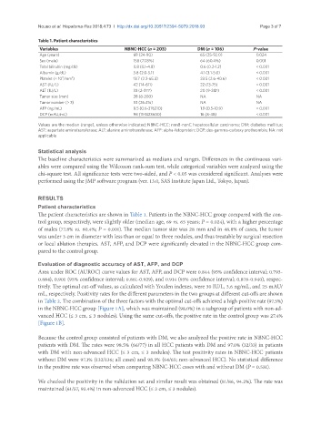
Nouso et al. Hepatoma Res 2018;4:73  I  http://dx.doi.org/10.20517/2394-5079.2018.93                                               Page 3 of 7


               Table 1. Patient characteristics
                Variables                      NBNC-HCC (n = 203)         DM (n = 106)         P-value
                Age (years)                       69 (24-90)               65 (25-92.0)         0.024
                Sex (male)                        158 (77.8%)              64 (60.4%)           0.001
                Total bilirubin (mg/dL)           0.8 (0.1-4.8)            0.6 (0.2-1.2)        < 0.001
                Albumin (g/dL)                    3.8 (2.0-5.1)            4.1 (3.1-5.0)        < 0.001
                        4
                            3
                Platelet (× 10 /mm )              13.7 (2.2-65.3)          23.5 (2.6-40.6)      < 0.001
                AST (IU/L)                        42 (14-611)              22 (13-75)           < 0.001
                ALT (IU/L)                        33 (2-377)               20 (9-247)           < 0.001
                Tumor size (mm)                   28 (8-200)               NA                   NA
                Tumor number (> 3)                53 (26.4%)               NA                   NA
                AFP (ng/mL)                       8.5 (0.6-376210)         1.9 (0.5-10.9)       < 0.001
                DCP (mAU/mL)                      98 (11-1323600)          16 (8-48)            < 0.001
               Values are the median (range), unless otherwise indicated; NBNC-HCC: nonB-nonC hepatocellular carcinoma; DM: diabetes mellitus;
               AST: aspartate aminotransferase; ALT: alanine aminotransferase; AFP: alpha-fetoprotein; DCP: des-gamma-carboxy prothrombin; NA: not
               applicable

               Statistical analysis
               The baseline characteristics were summarized as medians and ranges. Differences in the continuous vari-
               ables were compared using the Wilcoxon rank-sum test, while categorical variables were analyzed using the
               chi-square test. All significance tests were two-sided, and P < 0.05 was considered significant. Analyses were
               performed using the JMP software program (ver. 13.0, SAS Institute Japan Ltd., Tokyo, Japan).


               RESULTS
               Patient characteristics
               The patient characteristics are shown in Table 1. Patients in the NBNC-HCC group compared with the con-
               trol group, respectively, were slightly older (median age, 69 vs. 65 years; P = 0.024), with a higher percentage
               of males (77.8% vs. 60.4%; P = 0.001). The median tumor size was 28 mm and in 48.8% of cases, the tumor
               was under 3 cm in diameter with less than or equal to three nodules, and thus treatable by surgical resection
               or local ablation therapies. AST, AFP, and DCP were significantly elevated in the NBNC-HCC group com-
               pared to the control group.

               Evaluation of diagnostic accuracy of AST, AFP, and DCP
               Area under ROC (AUROC) curve values for AST, AFP, and DCP were 0.844 (95% confidence interval; 0.793-
               0.884), 0.901 (95% confidence interval; 0.861-0.929), and 0.914 (95% confidence interval; 0.878-0.940), respec-
               tively. The optimal cut-off values, as calculated with Youden indexes, were 30 IU/L, 3.6 ng/mL, and 25 mAU/
               mL, respectively. Positivity rates for the different parameters in the two groups at different cut-offs are shown
               in Table 2. The combination of the three factors with the optimal cut-offs achieved a high positive rate (97.5%)
               in the NBNC-HCC group [Figure 1A], which was maintained (98.0%) in a subgroup of patients with non-ad-
               vanced HCC (≤ 3 cm, ≤ 3 nodules). Using the same cut-offs, the positive rate in the control group was 27.4%
               [Figure 1B].

               Because the control group consisted of patients with DM, we also analyzed the positive rate in NBNC-HCC
               patients with DM. The rates were 98.5% (66/77) in all HCC patients with DM and 97.0% (32/33) in patients
               with DM with non-advanced HCC (≤ 3 cm, ≤ 3 nodules). The test positivity rates in NBNC-HCC patients
               without DM were 97.1% (132/136; all cases) and 98.5% (64/65; non-advanced HCC). No statistical difference
               in the positive rate was observed when comparing NBNC-HCC cases with and without DM (P = 0.531).

               We checked the positivity in the validation set and similar result was obtained (81/86, 94.2%). The rate was
               maintained (61/57, 93.4%) in non-advanced HCC (≤ 3 cm, ≤ 3 nodules).