
Page 73 - Read Online
P. 73
Ruff et al. Hepatoma Res 2023;9:37 https://dx.doi.org/10.20517/2394-5079.2023.51 Page 3 of 10
[18]
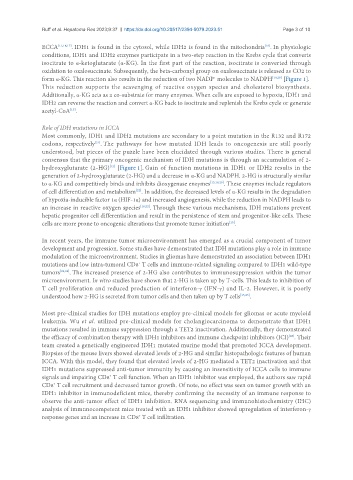
ECCA [11,16,17] . IDH1 is found in the cytosol, while IDH2 is found in the mitochondria . In physiologic
conditions, IDH1 and IDH2 enzymes participate in a two-step reaction in the Krebs cycle that converts
isocitrate to α-ketoglutarate (α-KG). In the first part of the reaction, isocitrate is converted through
oxidation to oxalosuccinate. Subsequently, the beta-carbonyl group on oxalosuccinate is released as CO2 to
form α-KG. This reaction also results in the reduction of two NADP molecules to NADPH [19,20] [Figure 1].
+
This reduction supports the scavenging of reactive oxygen species and cholesterol biosynthesis.
Additionally, α-KG acts as a co-substrate for many enzymes. When cells are exposed to hypoxia, IDH1 and
IDH2 can reverse the reaction and convert α-KG back to isocitrate and replenish the Krebs cycle or generate
acetyl-CoA .
[15]
Role of IDH mutations in ICCA
Most commonly, IDH1 and IDH2 mutations are secondary to a point mutation in the R132 and R172
codons, respectively . The pathways for how mutated IDH leads to oncogenesis are still poorly
[21]
understood, but pieces of the puzzle have been elucidated through various studies. There is general
consensus that the primary oncogenic mechanism of IDH mutations is through an accumulation of 2-
[15]
hydroxyglutarate (2-HG) [Figure 1]. Gain of function mutations in IDH1 or IDH2 results in the
generation of 2-hydroxyglutarate (2-HG) and a decrease in α-KG and NADPH. 2-HG is structurally similar
to α-KG and competitively binds and inhibits dioxygenase enzymes [15,18,20] . These enzymes include regulators
[22]
of cell differentiation and metabolism . In addition, the decreased levels of α-KG results in the degradation
of hypoxia-inducible factor 1α (HIF-1α) and increased angiogenesis, while the reduction in NADPH leads to
an increase in reactive oxygen species [18,22] . Through these various mechanisms, IDH mutations prevent
hepatic progenitor cell differentiation and result in the persistence of stem and progenitor-like cells. These
[15]
cells are more prone to oncogenic alterations that promote tumor initiation .
In recent years, the immune tumor microenvironment has emerged as a crucial component of tumor
development and progression. Some studies have demonstrated that IDH mutations play a role in immune
modulation of the microenvironment. Studies in gliomas have demonstrated an association between IDH1
mutations and low intra-tumoral CD8 T cells and immune-related signaling compared to IDH1 wild-type
+
tumors [23,24] . The increased presence of 2-HG also contributes to immunosuppression within the tumor
microenvironment. In vitro studies have shown that 2-HG is taken up by T-cells. This leads to inhibition of
T cell proliferation and reduced production of interferon-γ (IFN-γ) and IL-2. However, it is poorly
understood how 2-HG is secreted from tumor cells and then taken up by T cells [15,25] .
Most pre-clinical studies for IDH mutations employ pre-clinical models for gliomas or acute myeloid
leukemia. Wu et al. utilized pre-clinical models for cholangiocarcinoma to demonstrate that IDH1
mutations resulted in immune suppression through a TET2 inactivation. Additionally, they demonstrated
the efficacy of combination therapy with IDH1 inhibitors and immune checkpoint inhibitors (ICI) . Their
[26]
team created a genetically engineered IDH1 mutated murine model that promoted ICCA development.
Biopsies of the mouse livers showed elevated levels of 2-HG and similar histopathologic features of human
ICCA. With this model, they found that elevated levels of 2-HG mediated a TET2 inactivation and that
IDH1 mutations suppressed anti-tumor immunity by causing an insensitivity of ICCA cells to immune
signals and impairing CD8 T cell function. When an IDH1 inhibitor was employed, the authors saw rapid
+
CD8 T cell recruitment and decreased tumor growth. Of note, no effect was seen on tumor growth with an
+
IDH1 inhibitor in immunodeficient mice, thereby confirming the necessity of an immune response to
observe the anti-tumor effect of IDH1 inhibition. RNA sequencing and immunohistochemistry (IHC)
analysis of immunocompetent mice treated with an IDH1 inhibitor showed upregulation of interferon-γ
response genes and an increase in CD8 T cell infiltration.
+