
Page 64 - Read Online
P. 64
Page 367 Ganesan et al. Art Int Surg 2024;4:364-75 https://dx.doi.org/10.20517/ais.2024.68
Table 1. Summary of AI advancements in wound diagnosis
Study AI framework and/or computational system Outcome
Identification and classification
[11]
Birkner et al. Deep convolutional neural network trained with images Differentiate pyoderma gangrenosum from conventional leg
of pyoderma gangrenosum and leg ulcers ulcer
[12]
Hüsers et al. Image detection and classification algorithms of the Identify and classify venous leg ulcers and diabetic foot
YOLOv5, trained with 885 images of either wound ulcers
[13]
Swerdlow et al. and Convolutional neural network Segmentation and classification of pressure injury images
[14]
Zahia et al.
[15]
Chang et al. Deep learning based on superpixel segmentation Pressure ulcer diagnosis
[16]
Eldem et al. “Alexnet architecture”, a deep learning tool Classify pressure and diabetic wound images
Lau et al. [18] Smartphone application using a deep learning-based Detection and stage classification of printed images of
object detection system pressure injury wounds
Sizing
[17]
Mohammed et al. “Swift”, a noninvasive digital tool using AI Capture color calibrated images to identify wound
boundaries, surface area, and depth
[19]
Chan et al. Mobile device application using YOLOv4, validated with Detect length, width, and area of diabetic foot ulcers
144 photos
Tissue identification
Aldoulah et al. [20] SEEN-B4 deep learning framework Assess erythematous regions compared to an eschar or dry
crust
Veredas et al. [21] Neural networks and Bayesian classifiers Identify tissue types in wound images
[22]
Lien et al. Neural network model trained with three rounds of active Detect the growth of granulation tissue in diabetic foot
learning ulcers
[23]
Liu et al. EfficientNet deep learning model Create color-coded regions to identify ischemia and
Viswanathan et al. [24] AI-enabled noninvasive device, Illuminate®, capable of infection based on real patient images of diabetic foot ulcers
autofluorescence imaging
AI: Artificial intelligence; SEEN-B4: Swish-ELU EfficientNet-B4.
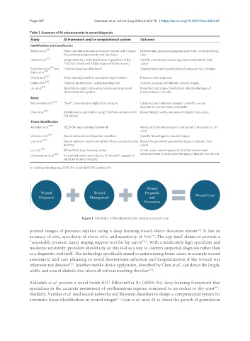
Figure 1. Schematic of the elements that comprise wound care.
printed images of pressure injuries using a deep learning-based object detection system . It has an
[18]
accuracy of 63%, specificity of above 85%, and sensitivity of 73% . The app itself claims to provide a
[18]
“reasonable pressure injury staging support tool for lay carers” . With a moderately high specificity and
[18]
moderate sensitivity, providers should rely on this tool as a way to confirm suspected diagnosis rather than
as a diagnostic tool itself. The technology specifically aimed to assist nursing home carers in accurate wound
assessment and care planning to avoid downstream infection and hospitalization if the wound was
otherwise not detected . Another mobile device application, described by Chan et al., can detect the length,
[18]
[19]
width, and area of diabetic foot ulcers all without touching the ulcer .
Aldoulah et al. present a novel Swish-ELU EfficientNet-B4 (SEEN-B4) deep learning framework that
[20]
specializes in the accurate assessment of erythematous regions compared to an eschar or dry crust .
Similarly, Veredas et al. used neural networks and Bayesian classifiers to design a computational system for
automatic tissue identification in wound images . Lien et al. used AI to detect the growth of granulation
[21]